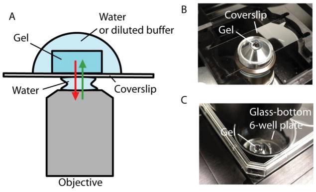
Figure 10.
Imaging expanded ExM samples with inverted microscopes. (A) Schematics showing the configuration of the objective and the expanded sample on an inverted microscope (with a water-immersion objective). The green and red arrows show the excitation and the emission light, respectively. (B) Typical imaging setup on an inverted spinning disk confocal microscope with a coverslip. (C) Typical imaging setup on an inverted spinning disk confocal microscope with a glass-bottom multi-well plate.

