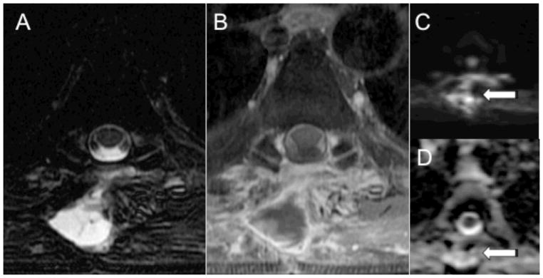
Figure 2.
MRI with DWI of the thoracic spine in a 65-year-old male with history of liver and renal transplant who presented with back pain after removal of thoracic masses. Axial T2 and post-gadolinium T1-weighted MR images (A, B) demonstrate a rim enhancing fluid collection in the paraspinal tissues at T5. DWI (C) demonstrates high intensity within the paraspinal collection (arrow) with corresponding high ADC (D, arrow). The ADC value measures 3268 × 10−6 mm2/sec. Microbiology of the fluid collection was negative.

