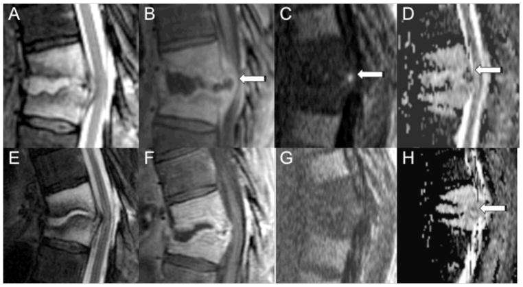
Figure 4.
MRI with DWI of the thoracic spine in a 64-year-old male with a history of thoracic radiation for esophageal cancer who presented with back pain before (A–D) and after four weeks of antibiotic therapy (E–H). Sagittal MR images demonstrate abnormal T2 hyperintensity (A, E) and enhancement (B, F) involving the T9 and T10 vertebral bodies with a rim enhancing fluid collection involving the T9-T10 disc extending into the epidural space (arrow). Before antibiotic therapy, DWI (C) shows focal hyperintensity within the ventral epidural space with associated low ADC (D) of 200 × 10−6 mm2/sec. Microbiology of the epidural fluid collection yielded Viridans group streptococci. DWI (G) after four weeks of antibiotic therapy shows resolution of the focal hyperintensity, and the ADC (H) is now relatively elevated (1556 × 10−6 mm2/sec).

