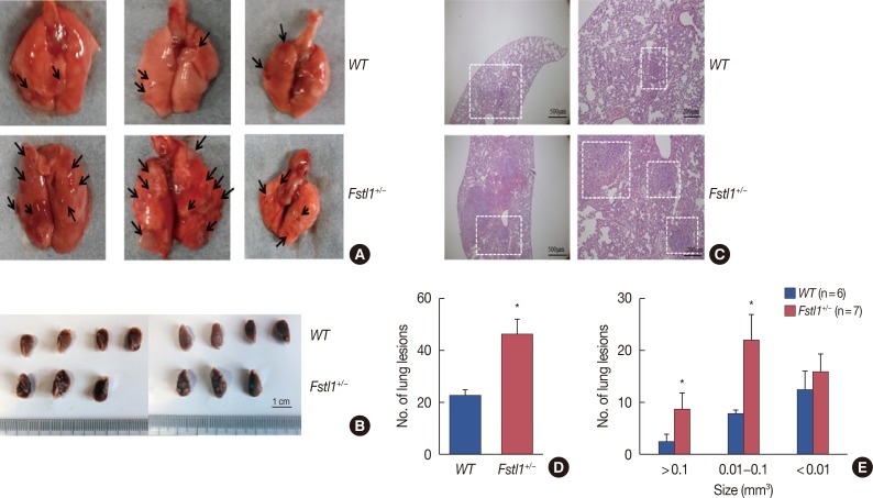
Figure 4. Increased lung metastasis in Fstl1+/−mice. (A) Thirty-one days after injection of 4T1 cancer cells, the mice were euthanized and analyzed for lung metastasis. The arrows were pointed to the lung metastasis in fresh lung tissue. (B) Thirty-one days after injection of 4T1 cancer cells, the mice were euthanized and analyzed for lung metastasis of formalin fixation. (C) Representative H&E stained images of the lung. (D) Number of lung metastatic foci were assessed in wild type (WT) (n=6) and Fstl1+/−mice (n=7). (E) Number and size of lung metastatic foci were assessed in WT (n=6) and Fstl1+/−mice (n=7). Each bar represents mean±SD for triplicate experiments.
*p<0.05, unpaired t-test.

