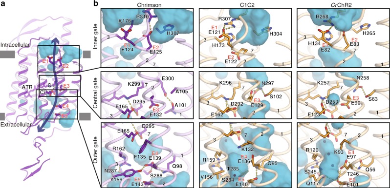
Fig. 2.
Ion pores of Chrimson and ChRs. a Water accessible cavities are illustrated in the Chrimson structure, with the putative ion pathway indicated by an arrow. Five glutamic acid residues lining the ion pore (E1–5) and two counterion residues (Ci1 and Ci2) are indicated by sticks, and the three constriction sites for the inner, central and outer gates are indicated by boxes. b Comparison of the constriction sites of Chrimson (left panels), C1C2 (center panels), and CrChR2 (right panels), for the inner (upper panels), central (middle panels), and outer (lower panels) gates. The constituent residues are shown as sticks, and the TM helix number is indicated on each helix

