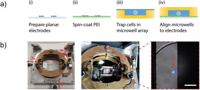
Figure 2.
Microwell arrays and patterned electrodes. (a) Microelectrodes with an interdigitated geometry are fabricated using the vacuum filling method. A layer of the polymer PEI is spun coat onto the electrodes to promote adhesion and prevent lift-off coating (1% w/v, 2500 rpm). A microwell array (14 × 9; well dimensions: 300 μm diameter, 50 μm height) is air-plasma treated and wetted with buffer solution before sedimenting a solution of cells. The microwell array containing cells is aligned to the interdigitated microelectrode. (b) A custom-built XYZ stage aids alignment. The inset composite image shows a nuclear stained (blue), GFP-expressing (green) cell trapped in a single microwell between two microelectrodes. Scale bar 100 μm.

