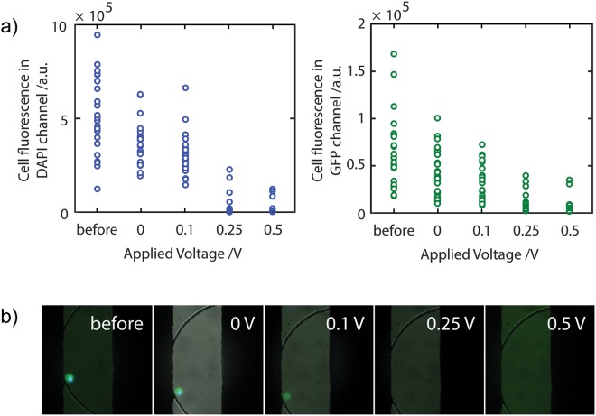
Figure 3.
Electrical lysis of cells. (a) Using the electrodes, an electric field is applied to the cells within the microwells. An alternating voltage Vac = 10 Vpp 1 MHz, superimposed to a constant DC voltage, VDC was applied. The GFP (left plot) and nuclear stain (right plot) fluorescence signal per cell was measured after 5 minute application of the electric field. (b) Combined bright field/GFP/H33342 images showing the effects of the electrical field on a cell at each of the DC voltage offsets indicated.

