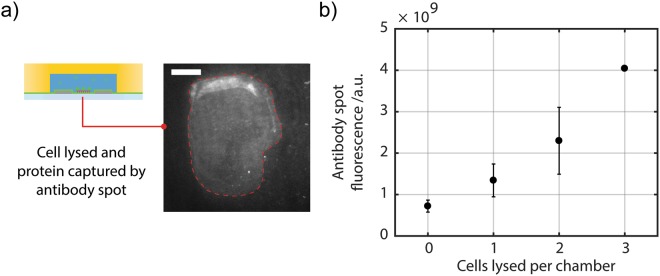
Figure 4.
Using microelectrodes in microwells to assay protein from cells. (a) The image, acquired by TIRF, shows an anti-GFP antibody spot printed onto a PEI- and microelectrode-coated coverslip and aligned into an individual well of the microwell array. Image shows GFP captured by the antibody spot (within dotted red line). Scale bar length is 25 μm. (b) The number of cells per well follows a Poisson distribution. The antibody spot in each microwell captures fluorescent GFP protein from the electrically lysed cells. The results show that, as expected, the GFP signal increases with increasing GFP-expressing cell occupancy.

