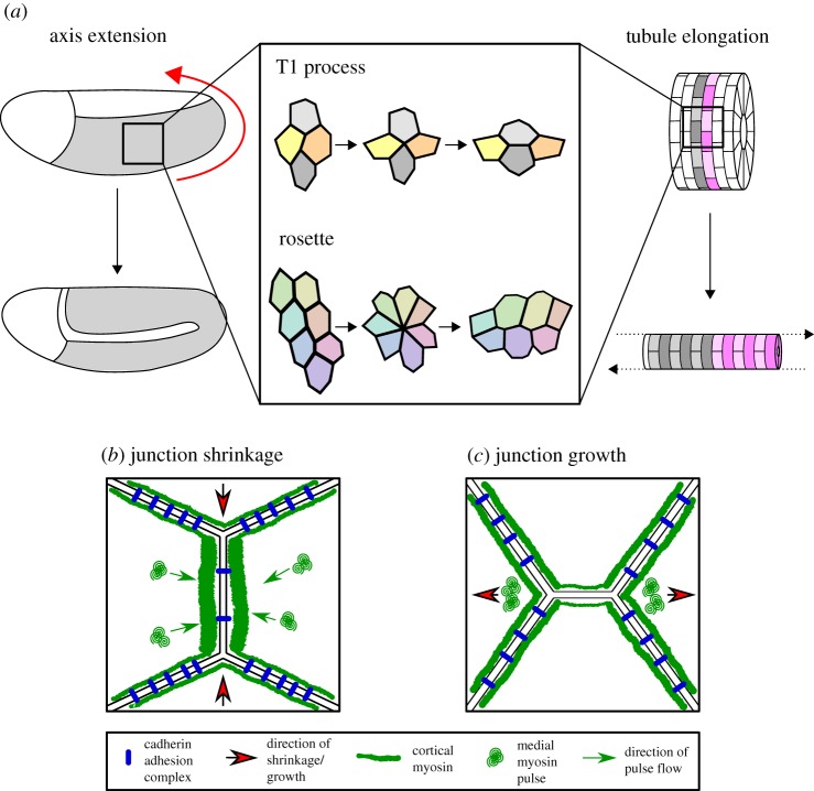
Figure 2.
Polarized intercalation deforms tissues during morphogenesis. (a) Morphogenesis, particularly examples of convergent extension such as axis extension (here shown Drosophila GBE, germband in grey, direction of elongation shown by red arrow) and tubule elongation, is often driven by polarized cell intercalation. Intercalation can take the form of either a T1 process in a tetrad of cells or the formation and resolution of a multicellular rosette. In Drosophila, junction shrinkage (b) is driven by planar polarized distributions of myosin II and cadherin adhesion complexes. Cortical junctional myosin is enriched at DV-oriented shrinking junctions, while cadherin adhesion complexes are enriched at stable AP-oriented junctions. Junction shrinkage is further driven by pulsatile flows of medial myosin, which flow into shrinking junctions. To achieve new junction growth (c), junctional myosin II activity must be reduced in the growing junction. Junctions then grow owing to cell non-autonomous forces generated by medial myosin pulses in adjacent cells, close to the ends of the new junction.

