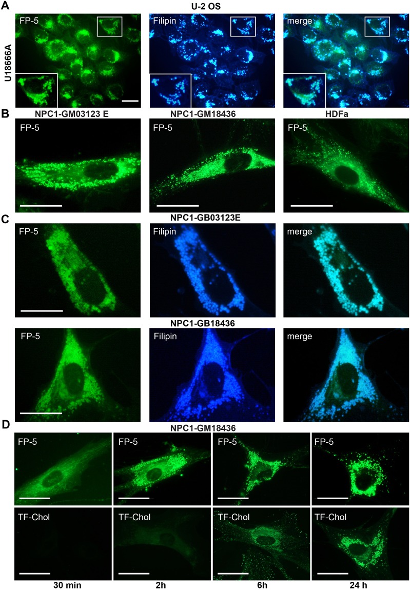
Figure 4.
FP-5 fluorescence in cells with abnormal content of cholesterol. (A) Cholesterol transport in U-2 OS was inhibited by inhibitor U18666A (1 μg/ml) for 48 h and then cells were labelled with FP-5 (200 nM) for additional 24 h, fixed and stained with filipin (50 μg/ml). Expansion of the region indicated by the white box is shown on the low left side. (B) Human fibroblasts carrying mutations in NPC1 cholesterol transporter (clones GB03123E, GB18436) and control normal human fibroblasts (HDFa) were labelled with FP-5 (200 nM) for 6 h and examined. (C) Co-localization of FP-5 and filipin staining in mutant cell clones. (D) Differential kinetics of FP-5 and TF-Chol lysosomal labelling in NPC-GM18436 fibroblasts. Cells were incubated with FP-5 (200 nM) and TF-Chol (1 μM) for indicated times in medium containing 5% LPDS and imaged live. Scale bar represents 10 μM.

