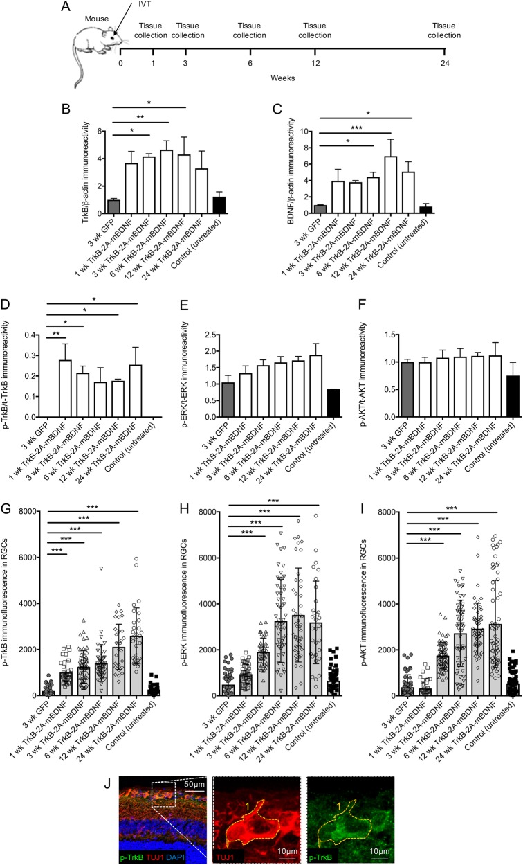
Fig. 4. Long-term vector expression and signaling in the mouse retina after intravitreal injection of AAV2 GFP or AAV2 TrkB-2A-mBDNF (2 µl, 1 × 10¹⁰ vector particles/eye).
a Schematic of the procedure performed and the time points in which tissues were collected. b, c Increased TrkB and BDNF expression was detected in retinal lysates at multiple time points post injection of AAV2 TrkB-2A-mBDNF compared to AAV2 GFP (n = 3/time point). d Increased p-TrkB was measured in retinal lysates expressing AAV2 TrkB-2A-mBDNF compared to AAV2 GFP or untreated controls (n = 3/time point). e, f Activated ERK (p-ERK) and AKT (p-AKT) showed a trend for increased expression following transduction with AAV2 TrkB-2A-mBDNF when assessed in whole retinal lysates (n = 3/time point). g–i p-TrkB, p-ERK, and p-AKT immunofluorescence within individual RGCs (TUJ1+ cells) was significantly increased in eyes injected with AAV2 TrkB-2A-mBDNF compared to controls (n = 2000 RGCs from 14 retinas). j Representative image showing how immunofluorescence was measured within individual RGCs (TUJ1+ cells). *P < 0.05, **P < 0.01 and ***P < 0.001 compared to 3-week GFP. Graphs show mean ± SEM with P values obtained via a one-way ANOVA followed by Bonferroni-modified t-tests for multiple comparisons

