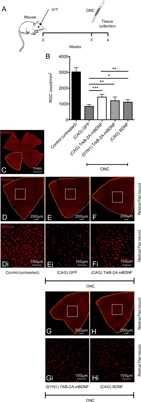
Fig. 6. Neuroprotection using AAV2 TrkB-2A-mBDNF vs AAV2 BDNF in a mouse optic nerve crush (ONC) injury model.

a Schematic of the procedure performed. Vectors were administered 3 weeks prior to ONC (2 µl, 9.14 × 10⁹ vector particles/eye). b RGC (Brn3A+ cells) were counted 1 week after ONC, 8 images quantified/retina. c Representative image of an entire mouse retinal flat mount. d–hi Representative image of a retinal quadrant showing the size of a single zoomed in image (white box) which was quantified and used to estimate RGC number. Control (untreated) n = 10, (CAG) GFP n = 9, (CAG) TrkB-2A-mBDNF n = 7, (SYN1) TrkB-2A-mBDNF n = 6, (CAG) BDNF n = 7. *P < 0.05, **P < 0.01 and ***P < 0.001 compared to AAV2 GFP transduced eyes which had ONC. The graph shows mean ± SD with P values obtained via a one-way ANOVA followed by Bonferroni-modified t-tests for multiple comparisons. CAG = cytomegalovirus/chicken beta-actin hybrid promoter, SYN1 = synapsin-1 promoter
