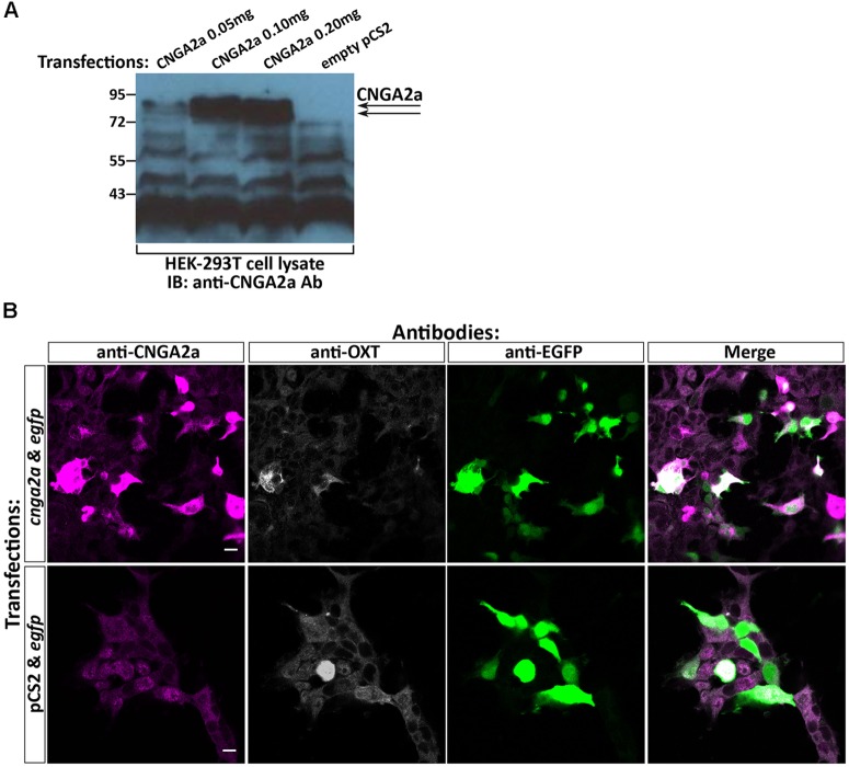
FIGURE 2.
Specificity of anti-CNGA2a mAb in heterologous expression system. (A) Western blot analysis of HEK293T cells transfected with cnga2a cDNA. HEK293T cells were transiently transfected with different amounts of cnga2a cDNA or a mock plasmid and were harvested 48 h post-transfection. Western blot analysis of equal amounts of protein extracts were performed using anti-CNGA2a mAb. The correct position of the doublet CNGA2a protein bands are marked by arrowheads (n = 2/2). (B) Confocal images of HEK293T cells transfected with cnga2a cDNA. HEK293T cells were transiently co-transfected with combinations of egfp cDNA either with cnga2a cDNA or an empty pcS2 plasmid. Forty-eight hours post-transfection the monolayer cultures were fixed in 3% paraformaldehyde (PFA), permeabilized with 0.5% Triton-X100/3% PFA, washed in PBS and fluorescently co-stained with anti-CNGA2a (magenta), anti-OXT (gray scale), and anti-GFP (green) antibodies (n = 4/4). Scale bars: 10 μm.

