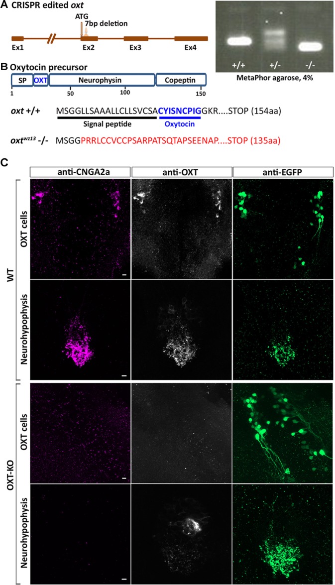
FIGURE 5.

Anti-CNGA2a immunoreactivity is lost following KO of oxytocin. (A) Schematic representation of oxytocin (oxt) gene. OXT translation start site and sgRNA target site are both indicated with arrows. Embryos and adult fish were screened by PCR for germline transmission using gene-specific primers following PCR products separation in 4% MetaPhor gels. DNA sequence of mutant allele transmitted through the germline contained a 7 bp deletion. (B) Schematic representation of predicted translated products from oxt +/+ and oxt –/– alleles. (C) Confocal images showing representative anti-CNGA2a (magenta) and anti-OXT (gray scale) labeling of OXT cells and their neurohypophyseal axonal termini of OXT-KO 6-day-old larva in the background of a transgenic Tg(oxt:egfp-3′utr) reporter (green) (n = 12/12). Scale bars: 10 μm.
