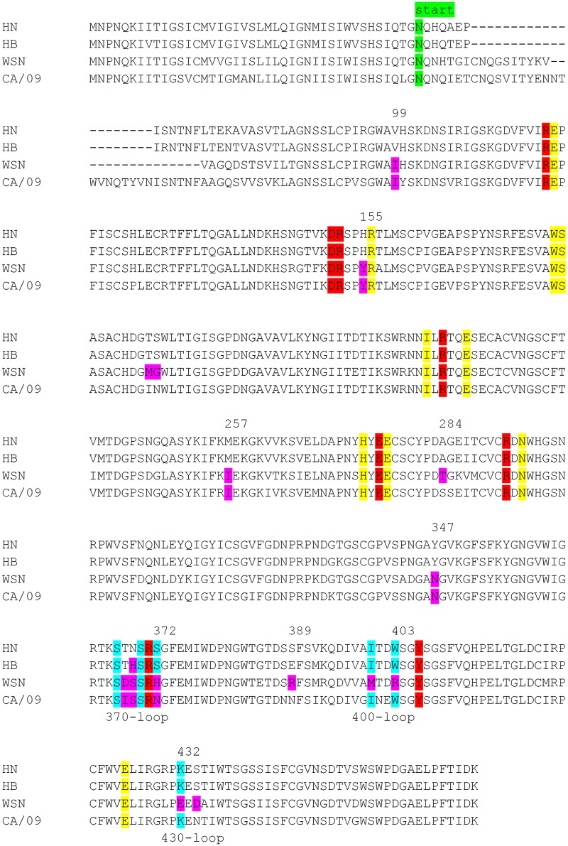
FIG 3.
Alignment of N1 proteins. Alignment of several N1 proteins analyzed in this study is shown. The start of the N1 protein ectodomain expressed as a recombinant soluble fusion protein is indicated. Catalytic and framework residues in the active site are shown in red and yellow, respectively (5, 6). SIA contact residues in the 2nd SIA-binding site, based on the N9 crystal structure (7), are shown in light blue. Residues introduced in HN N1 are shown in purple. N2 numbering is indicated.

