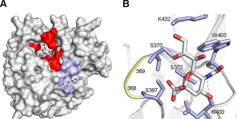
FIG 4.

Structure of the N9 2nd SIA-binding site. (A and B) Crystal structure of N9 from A/tern/Australia/G70C/75 in complex with SIA (N-acetylneuraminic acid) (PDB 1MWE) (7). (A) Surface representation. The NA active site and the 2nd SIA-binding site (SIA contact residues) are shown in red and light blue, respectively. The SIA moieties in these sites are shown as sticks (oxygen in red, nitrogen in blue, and carbon in gray). (B) Structure of the 2nd SIA-binding site. SIA is shown as sticks (oxygen in red, nitrogen in blue, and carbon in gray). Residues in the 2nd SIA-binding site that directly contact SIA (S367, S370, S372, N400, W403, and K432) are shown in stick representation (light blue), amino acids at positions 368 and 369 that differ between HN and WSN and CA/09 are shown in cartoon representation (yellow). Hydrogen bonds between SIA and residues in the 2nd SIA-binding site are shown as dashed red lines. Figures were made using PyMOL.
