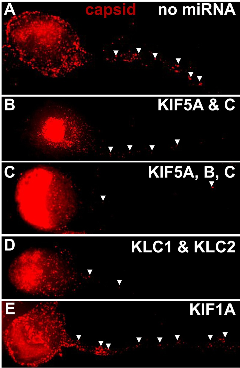
FIG 10.

Fluorescent imaging of HSV capsids in neurons expressing kinesin-specific miRNAs. CAD neurons were infected with baculoviruses expressing GFP and also no miRNA (A) and miRNAs targeting KIF5A and -5C (B), KIF5A, -5B, and -5C (C), KLC1 and KLC2 (D), or KIF1A (E) for 54 h. Subsequently, the neurons were infected for 21 h with HSV GS2822, which expresses VP26-RFP, and then fixed with 4% PFA. CAD neurons expressing strong GFP signal were selected for imaging of RFP-labeled capsids in axons, which are indicated by arrowheads. (A) CAD neurons that were transduced with a baculovirus expressing GFP but no miRNA (control) displayed numerous HSV capsids in axons. (B) Neurons expressing KIF5A- and -5C-specific miRNAs showed fewer capsids in axons than the control. (C) Neurons expressing KIF5A-, -5B-, and -5C-specific miRNAs showed further reduced HSV capsids in axons. (D) Neurons expressing KLC1- and KLC2-specific miRNAs also showed reduced HSV capsids in axons. (E) In contrast, neurons expressing KIF1A-specific miRNAs showed only a small reduction in capsid numbers in axons.
