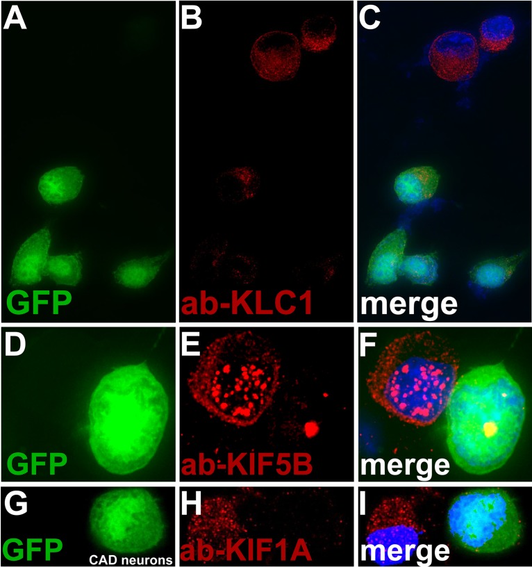
FIG 9.
Fluorescent imaging of kinesins present in axons following expression of miRNAs targeting kinesins. CAD neurons were infected with baculoviruses expressing miRNAs targeting kinesin-1 light chains KLC1 and KLC2, as well as GFP (A to C), miRNAs targeting kinesin-1 heavy chains KIF5A, -5B, and -5C and GFP (D to F), and miRNAs targeting kinesin-1 KIF1A and GFP (G to I). After 72 h, the neurons were fixed with 4% PFA, permeabilized, and immunostained with antibodies specific for KLC1 (A to C), KIF5B (D to F), or KIF1A (G to I) and also with 300 nM DAPI. CAD neurons with strong GFP expression exhibited little KLC1 (red) (A to C), little KIF5B (D to F), and little KIF1A (G to I).

