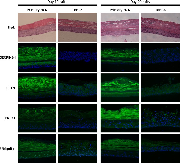
FIG 6.
Immunofluorescence staining of raft tissue. The spatial protein expression of SERPINB4, RPTN, KRT23, and ubiquitin was observed by performing immunofluorescence staining of primary HCK and 16HCK raft tissue fixed at 10 days and 20 days of culture (green, target protein; blue, nuclear staining). For experiments with day 10 raft tissues, three raft experiments, each with 16HCK and primary HCK cell lines representing three donors each, were set up. For experiments with day 20 raft tissues, six raft experiments, each with 16HCK and primary HCK cell lines representing three and six donors, respectively, were set up. Magnifications, ×200. H&E, hematoxylin and eosin staining. Images were acquired with a Nikon Eclipse 80i microscope and NIS Elements (version 4.4) software.

