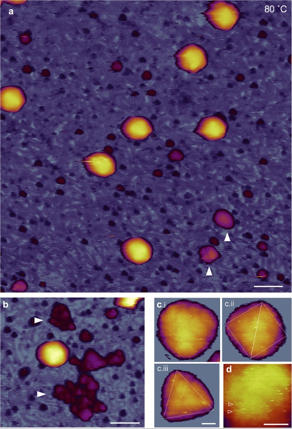
FIG 6.

AFM of T7 phages treated at 80°C. (a) Overview of a 1-μm by 1-μm sample area. White arrowheads point at large (>10 nm) globular particles. Scale bar, 100 nm. (b) AFM image showing large aggregates of globular particles (white arrowheads) (c) High-resolution AFM images of 80-degree-treated T7 phage particles with resolvable capsomeres on their surface. Views along the 3-fold (i and iii) and 2-fold (ii) symmetry axes are shown. Scale bar, 10 nm. (d) Magnified view of the capsomeric structure. Arrowheads point at putative spokes of the originally cogwheel-shaped capsomere. Note that the central pore cannot be resolved, most likely due to the swelling of the protein matrix. Scale bar, 10 nm.
