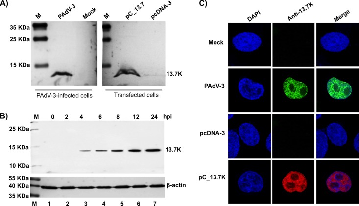
FIG 1.
Expression of 13.7K. (A, B) Western blots. Proteins from the lysates of mock-infected cells (lanes 1), PAdV-3-infected cells (panel A, lane 2; panel B, lanes 2 to 7), plasmid pcDNA-3 DNA-transfected cells (panel A, lane 3), or plasmid pC_13.7 DNA-transfected VIDO R1 cells (6) (panel A, lane 4) were separated by 12% SDS-PAGE, transferred to nitrocellulose, and analyzed by Western blotting using anti-13.7K serum. Expression of β-actin using an anti-β-actin MAb was used as a loading control. The positions of molecular weight markers in kilodaltons are shown on the left. The molecular weights (in kilodaltons) of observed proteins are shown on the right. (C) Subcellular localization of PAdV-3 13.7K. Monolayers of VIDO R1 (6) cells mock infected (top row), infected with PAdV-3 (second row), transfected with plasmid pcDNA-3 DNA (third row), or transfected with plasmid pC_13.7K DNA (bottom row) were analyzed by indirect immunofluorescence using anti 13.7K serum and Alexa Fluor 488-conjugated (top and second rows) or TRITC-conjugated (third and bottom rows) goat anti-rabbit serum. The cells were mounted in a Vectashield mounting medium with DAPI (Vector Laboratories) to stain nuclei and monitored by confocal microscope.

