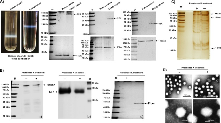
FIG 2.
Analysis of CsCl-purified PAdV-3 virions. (A) CsCl density gradient purification of PAdV-3. The upper band contains empty capsids (EC), and the lower band contains mature capsid (MC) (upper panels). Two to five micrograms of PAdV-3 empty capsids or mature capsids was used with anti-13.7K, anti-52K (23), antifiber (23), antihexon (23) anti-22K (23), or anti-33K (23) serum. (B) Proteinase K treatment. Proteins from purified PAdV-3 untreated (lane −) or treated (lane +) with 20 μg of proteinase K were separated by 10% to 12% SDS-PAGE, transferred to nitrocellulose, and probed with antihexon serum (a), anti-13.7K serum (b), and antifiber serum (c). The molecular weight markers (M) in kilodaltons are shown. (C) Silver staining. Proteins from purified PAdV-3 untreated (lane −) or treated (lane +) with 20 μg of proteinase K were separated by 12% SDS-PAGE, and the gel was stained with silver staining. The positions of the molecular weights (M) in kilodaltons are shown on the left. (D) Transmission electron microscopy. Purified PAdV-3 virions untreated (lane −) or treated (lane +) with 20 μg proteinase K were negatively stained with 2% aqueous phosphotungstic acid and analyzed by transmission electron microscopy.

