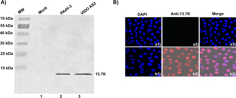
FIG 4.
Expression of 13.7K in VIDO AS2 cell line. (A) Western blot. Proteins from the lysates (10 μl) of VIDO R1 cells (6) mock infected (lane 1) or infected with PAdV-3 at an MOI of 2 (lane 2) and of VIDO AS2 cells (lane 3) were analyzed by Western blotting using anti-13.7K serum. (B) Indirect immunofluorescence. Monolayers of VIDO-R1 cells (a1 to a3) or VIDO AS2 cells (expressing protein 13.7K) (b1 to b3) were fixed with 3.7% paraformaldehyde and visualized by indirect immunostaining using anti-13.7K serum followed by TRITC-conjugated goat anti-rabbit IgG using a confocal microscope. The nuclei were stained by DAPI.

