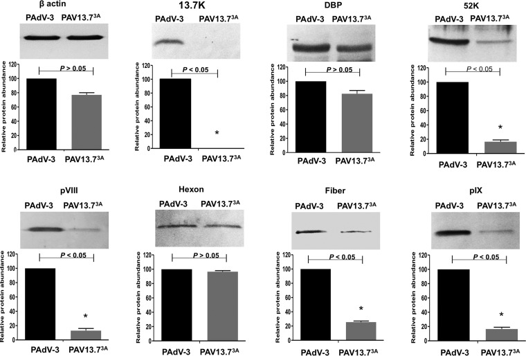
FIG 6.
Analysis of viral protein expression in virus-infected cells. Proteins from the lysates of PAdV-3- or PAV13.73A-infected cells were analyzed by Western blotting using anti-pIX (23), anti-DBP (30), anti-13.7K (this study), anti-52K (23), antihexon (23), anti-pVIII (50), and anti-fiber (22) antibodies followed by alkaline phosphatase-conjugated secondary antibodies. The β-actin was detected using anti-β-actin MAb followed by alkaline phosphatase-conjugated secondary antibodies. The BCIP/NBT solution was used as a substrate to visualize proteins. The results were quantified using ImageJ software (http://rsb.info.nih.gov/ij/). Values represent the means from two independent experiments, and error bars indicate SD. Significant differences (P < 0.05) are indicated with an asterisk (*).

