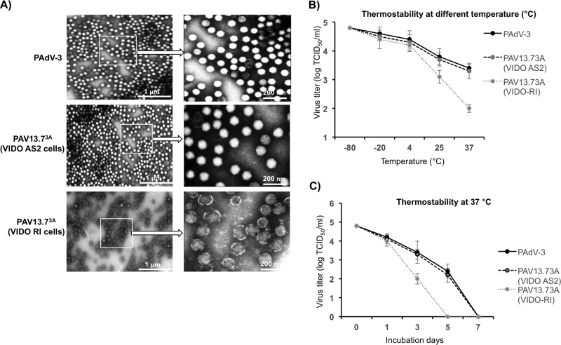
FIG 9.
Purified virus analysis and interaction of protein 13.7K with other PADV-3 protein(s). (A) Electron microscopic analysis. Purified PAdV-3 and PAV13.73A grown in VIDO AS2 cells and PAV13.73A grown in VIDO R-1 cells (6) are shown as indicated at a magnification of ×30,000. The arrows indicate enlargements of selected boxed regions of each virus (magnification, ×1,000,000). (B, C) Thermostability assay. Purified virions (105 TCID50) of PAdV-3 and PAV13.73A grown in either VIDO R1 or VIDO AS2 (expressing protein 13.7K) cells were incubated at different temperatures for 3 days, and the residual viral infectivity was determined by TCID50 (B). Purified virions (105 TCID50) of PAdV-3 and PAV13.73A grown in either VIDO R1 or VIDO AS2 cells were incubated at 37°C for the indicated time points (0, 1, 3, or 7 days postinfection), and the residual viral infectivity was determined by TCID50 (C).

