
FIG 4.
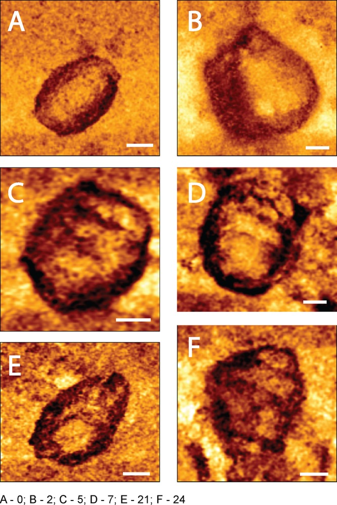
High-resolution spatial mechanical mapping of an HIV-1 core surface treated with 10 μM PF74 during reverse transcription. Cores were adhered to HMDS-coated glass slides and kept in MOPS buffer. Reactions were initiated by adding dNTPs and MgCl2 to the cores. Images were acquired using the QI mode. (A) The mechanical map of a core before the beginning of reverse transcription. (B to F) Mechanical maps of cores after 2, 5, 7, 21, and 24 h of reverse transcription, respectively. Scale bar, 50 nm.