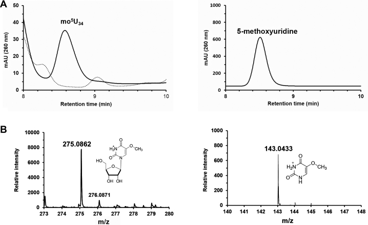
Figure 4.
In vitro mo5U forming activity of TrmR. (A) On the left, HPLC analysis of TrmR directed the formation of mo5U (solid line) versus control which contains no TrmR (dashed line). The peak corresponding mo5U eluted from the C-18 column at 8.6 min which has been confirmed by spiking with an authentic compound (right panel). (B) The assay sample was analyzed by LC-MS/MS in positive mode, where the peak corresponding to mo5U ([C10H15N2O7]+, calculated m/z = 275.088) was selected (left) and the subsequent fragmentation by ESI was confirmed as detection of 5-methoxyuracil ([C5H7N2O3]+, calculated m/z = 143.046) in MS/MS (right).

