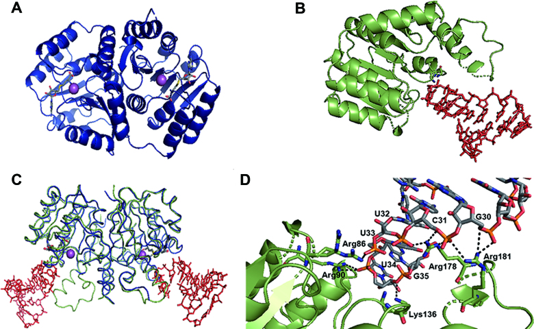
Figure 5.
Overall structures of TrmR. The ribbon presentation of the crystal structure of (A) RNA-free TrmR (navy) with SAH (gray stick) and magnesium ion (sphere, magenta), and (B) TrmR (green) bound to tRNAASL (red stick) with SAM (gray stick). (C) Overlaid structures of RNA-free TrmR (blue) and TrmR:tRNAASL (green and red). (D) Ionic/hydrogen bond interactions between ASL tRNA and a set of basic residues of TrmR in the crystal structure of TrmR–ASLAla complex. The ASL is shown with their carbon atoms colored gray, nitrogen atoms colored blue, oxygen atoms colored red and phosphorous atoms colored orange. Hydrogen bond is indicated by dashed lines.

