Figure 7.
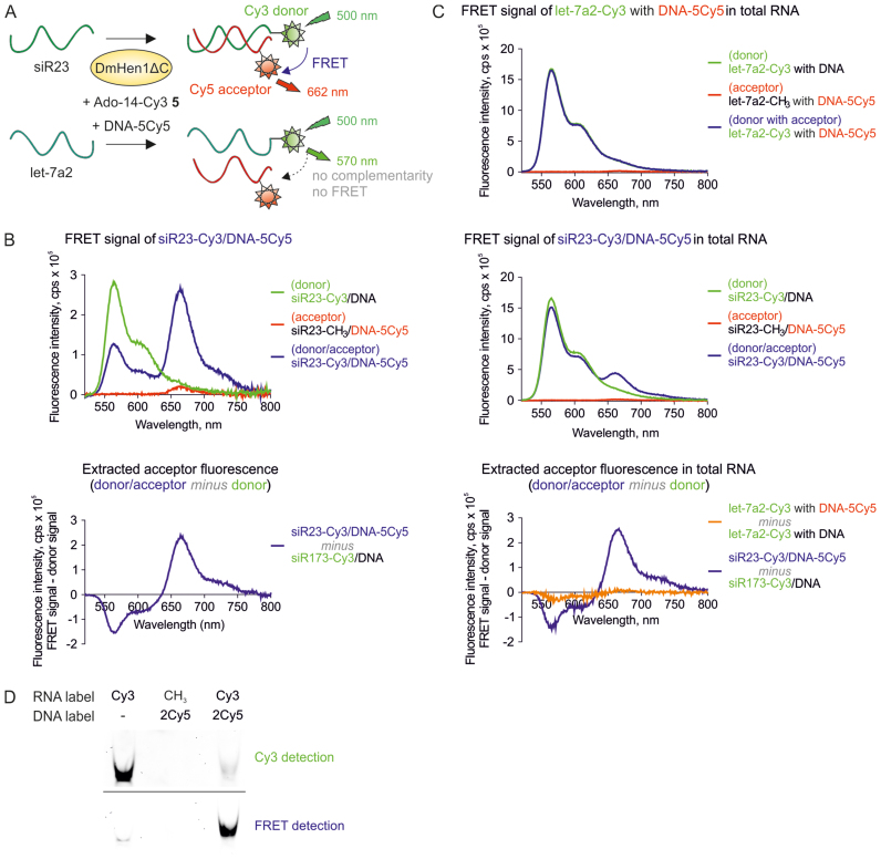
Selective detection of DmHen1-labelled ssRNA using FRET assay. (A) A schematic illustration of the experiment. ssRNA was 3′-end labelled in one step with Cy3 fluorophore transferred from Ado-14-Cy3 5 cofactor by DmHen1ΔC and later annealed to the siR23-complementary DNA bearing Cy5 at 5th position. The FRET signal at 662 nm appears only if Cy3-RNA donor and DNA-born Cy5 acceptor are close enough for an energy transfer. (B) Detection of FRET signal of Cy3/Cy5 pair in solution. siR23 Cy3-labelled by 1 μM of DmHen1ΔC in the presence of 3 μM of Ado-14-Cy3 was annealed to a complementary DNA in equimolar concentration. The top graph shows the emission spectra of siR23-Cy3/DNA-5Cy5 FRET pair excited at 500 nm and two control samples: donor siR23-Cy3/DNA containing RNA-born Cy3 and intact DNA and acceptor siR23-CH3/DNA-5Cy5-methylated RNA and DNA-born Cy5. The graph at the bottom highlights the extracted acceptor fluorescence acquired by subtracting the donor fluorescence from two-coloured spectra. (C) Specific detection of siR23 in total RNA sample through FRET signal registration. Top: FRET emission at 662 nm did not appear in Cy3-labelled total RNA with let-7a2, which was not complementary to DNA-5Cy5. Middle: the presence of siR23 in the sample is proved by emerging FRET signal. Bottom: extracted acceptor fluorescence at 662 nm is only visible upon addition of siR23. Samples were prepared as described earlier with the exception that 0.5 μg/μl of total RNA from HCT116 human colon carcinoma cell line was added in the labelling reaction and 10 μM of cofactor was used. (D) Visualization of siR23-Cy3/DNA-2Cy5 FRET pair in polyacrylamide gel. Cy3 and FRET signals were detected by exciting the fluorophores with green laser at 532 nm and registering their emission using LPG (575 nm) and LPR (665 nm) filters, respectively.

