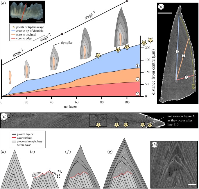
Figure 1.
Reconstruction of growth dynamics obtained from a histological thin section of Ozarkodina confluens. (a) Morphological change calibrated with individual growth layers (lamellae of the crown tissue). (b) Composite BSE images of polished thin section (scale bar 50 µm) through the conodont element outlining three transects along which lamellae were counted. (c) Episodes of wear recorded as tip breakages. (d–g) Reconstruction of wear followed by repair of the damaged element. (h) BSE image of wear surface (scale bar 5 µm). (Online version in colour.)

