Figure 3.
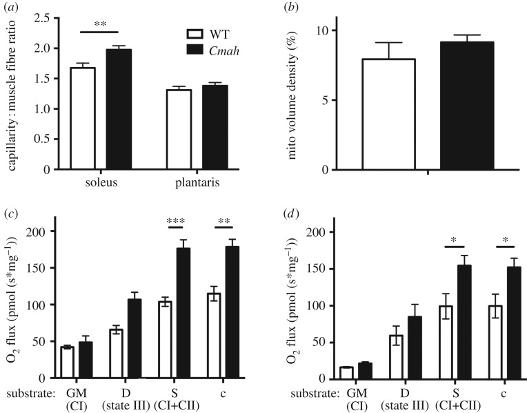
Skeletal muscle vascularity and mitochondrial respiration in WT and Cmah−/− mice. (a) Muscle capillary-to-fibre ratio was measured by lead ATPase stain in the soleus (n = 6) and plantaris (n = 5). (b) Mitochondrial volume density was also measured by transmission electron microscopy (TEM) of the tibialis anterior (n = 3). (c) Diaphragm (n = 4 WT and 6 Cmah−/−) and (d) soleus (n = 6) saponin-permeabilized muscle fibre bundles were exposed to saturating conditions of the following substrates: glutamate and malate (GM) to measure complex I respiration, ADP to measure state III respiration, complex II substrate succinate (Suc) to measure complex I and complex II respiration combined and cytochrome c (Cyt c) as a quality control to ensure mitochondrial outer membrane integrity. Error bars are s.e.m. Statistics were determined using two-way ANOVA with Tukey's multiple comparison test. *p < 0.05, **p < 0.01 and ***p < 0.001 represent estimates of statistical significance.

