Fig. 2.
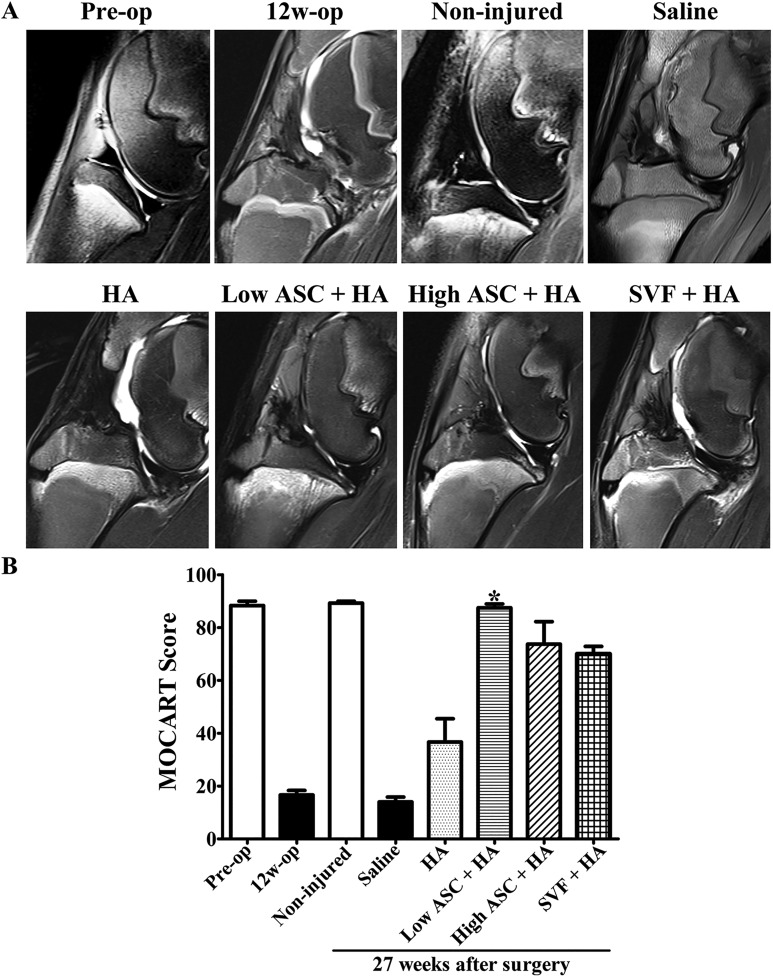
After OA model was successfully induced, MR imaging indicated the efficacy of autologous low-dose ASC+HA treatment. (A) MRI was performed at pre-operation (pre-op), 12 weeks post-operation (12 w), and 27 weeks post-operation in different treatments (non-injured, saline, HA, low ASC+HA, high ASC+HA, and SVF+HA). In the pre-op and non-injured groups, T2 sequence of 3.0 T MRI showed smooth cartilage layer and clear synovium lining; however, at 12 weeks post-surgery and in the saline and HA groups, the T2 sequence of the MRI exhibited damaged cartilage and hydropic synovium. ASC+HA and SVF+HA treatments improved cartilage injury, bone marrow lesions of subchondral bone, synovial hydropic swelling. (B) MOCART scores in low-dose ASC+HA treatment were significantly higher than those in the saline and HA groups. The P-values were obtained using a Kruskal–Wallis test and Dunn’s post-test (*P<0.05). Data were collected from three repetitions (n=3).
ASC: adipose mesenchymal stem cell; HA: hyaluronic acid; MOCART: magnetic resonance observation of cartilage repair tissue; MRI: magnetic resonance imaging; OA: osteoarthritis; SVF: stromal vascular fraction.

