Fig. 3.
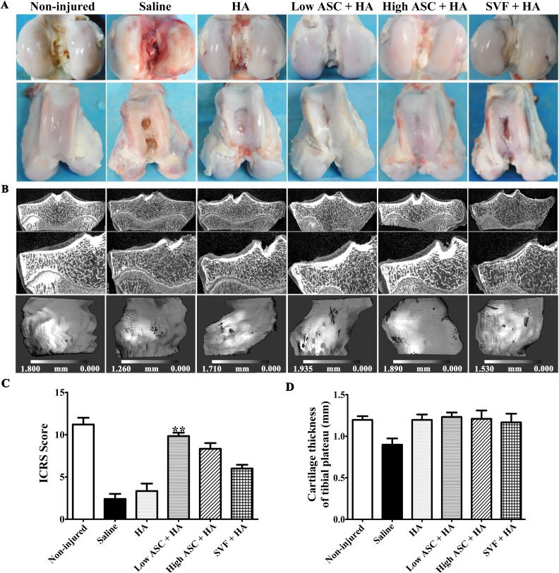
Macroscopic analysis and µCT scanning showed autologous ASC+HA exhibited better efficacy than SVF+HA in treating OA. (A) Macroscopy of the articular cartilage surface of femoral condyles at 27 weeks after surgery (12 weeks post second injection) showed more continuous and smoother cartilage layer with no obvious cartilage destruction in the ASC-treated groups than the SVF group. In contrast, visible fissures and cracks to different extent were observed in the saline and HA groups. (B) µCT evaluations displayed cartilage thickness of tibial plateau at 12 weeks post second injection in each group. After contouring out the underlying subchondral bone, three-dimensional images of the medial tibial condyle cartilage surface were reconstructed, respectively. The gray scale represents the thickness of the cartilage at each voxel. Dark areas indicate thinner articular cartilage, while light areas represent thicker articular cartilage. (C) Only low-dose ASC+HA treatment obtained higher ICRS scores of femoral condyles than those of the saline group. (D) Average cartilage thickness of the tibial plateau measured by µCT images showed that there were no differences in ASC+HA and SVF+HA-treated groups. The P-values were obtained using Kruskal–Wallis test and Dunn’s post-test (**P<0.01). Data were collected from three repetitions (n=3).
µCT: micro-computed tomography; ASC: adipose mesenchymal stem cell; HA: hyaluronic acid; ICRS: International Cartilage Research Society; OA: osteoarthritis; SVF: stromal vascular fraction.

