Fig. 5.
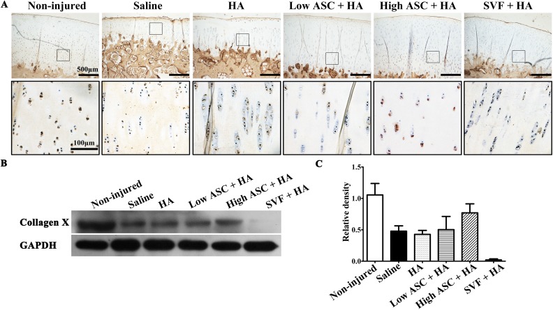
Type X collagen expression exhibited cartilage quality of autologous ASC+HA and SVF+HA treatments. (A) Immunohistochemistry results showed that hypertrophic chondrocytes expressing collagen X were in alignment at the deep of articular cartilage in the non-injured group. Few collagen-X-positive chondrocytes were observed in the saline and HA-treated groups while high-dose ASC+HA treatment rescued hypertrophic chondrocytes expressing collagen X at the deep area of articular cartilage. It seemed that low-dose ASC+HA treatment showed more collagen-X-positive hypertrophic chondrocytes than that of in SVF+HA treatment. (B, C) Western blot and densitometry analysis results also showed that high- and low-dose ASC+HA treatments expressed collagen X while SVF+HA treatment showed weak expression.
ASC: adipose mesenchymal stem cell; HA: hyaluronic acid; SVF: stromal vascular fraction.

