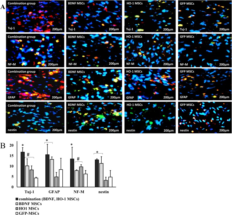
Figure 6.
Immunofluorescent staining for the expression of neural markers. (a) images of stained slides. The slides were stained with Tuj-1, NF-M, GFAP, and nestin as red, and the nucleus was stained with DAPI as blue. Each image represents the four samples per group with a scale bar of 200 μm. (b) Percentage expression of neural marker positive cells. The percentage of cells positive for Tuj-1 and NF-M was highest in the combination group, followed by higher percentages in BDNF- and HO-1-MSCs groups compared to the GFP-MSCs group (*#P ≤ 0.05). While the cells positive for GFAP and nestin were similar between combination and BDNF-MSCs groups, they were higher than the HO-1- and GFP-MSCs groups (*P ≤ 0.05).

