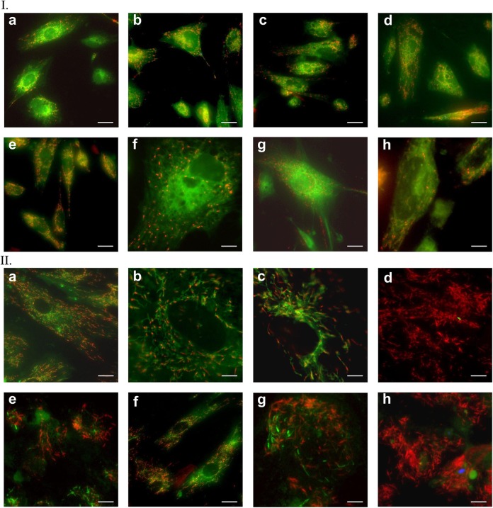
Figure 11.
Mitochondrial staining with JC-1 dye on the 20th (I) and 40th (II) day of SMiPSC-CM in vitro differentiation culture. (a) to (h) Pictures of both analyzed time points refer to selected stained areas of in vitro cell culture. Scale bar: 50 μm and 150 μm for II(g) and II(h) images, respectively.

