Figure 4.
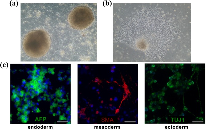
Images taken from spontaneous in vitro differentiation via embryoid bodies: (a) SMiPSC-derived embryoid bodies on day 5 of in vitro suspension culture; (b) outgrowing embryoid body in adherent cell culture; (c) immunolabeled EBs demonstrated α-fetoprotein (AFP), smooth muscle actin (SMA), and neural class III β-tubulin (TUJ-1) expression. Magnification of (a) and (b) pictures at 10×. Scale is 50 μm.

