
Figure 8.
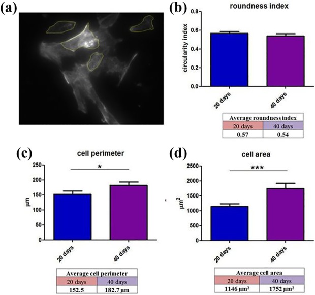
Morphological parameters measured in cardiac myocytes (n=70) in in vitro cell differentiation culture: (a) Image included for measurement with LAS X software. Calculations were given as follows: (b) roundness index, (c) cell perimeter, and (d) cell area. Plots: mean value + SEM.