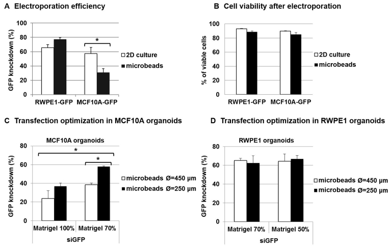
Figure 4.
Improving transfection efficiency by modulating microbead size and Matrigel concentration. RWPE-1-GFP and MCF10A-GFP-encapsulated organoids or 2D cell cultures were electroporated with siRNA (control siAllStars and siGFP, 20 nM). After 3 days, transfection efficiency (A) was determined using flow cytometry via GFP MFI measurement, as follows: transfection efficiency = 100 − (MFI siGFP/MFI siAllStars) × 100 and cell viability (B) was measured via trypan blue dye exclusion staining. The results represent the mean value ± SEM of three experiments (*P < 0.05). (C) Transfection optimization in MCF10A organoids: MCF10A-GFP microbeads of different sizes (450 and 250 μm in diameter Ø) and Matrigel concentrations (70 and 100%) were produced and electroporated with siRNA (control siAllStars and siGFP, 20 nM). After 3 days, extinction efficiency was determined using flow cytometry, as previously described. (D) Transfection optimization in RWPE1 organoids: RWPE-1-GFP microbeads of different sizes (450 and 250 μm in diameter Ø) and Matrigel concentrations (50 and 70%) were produced and electroporated with siRNA (control siAllStars and siGFP, 20 nM). After 3 days, extinction efficiency was determined using flow cytometry, as previously described.

