Figure 1.
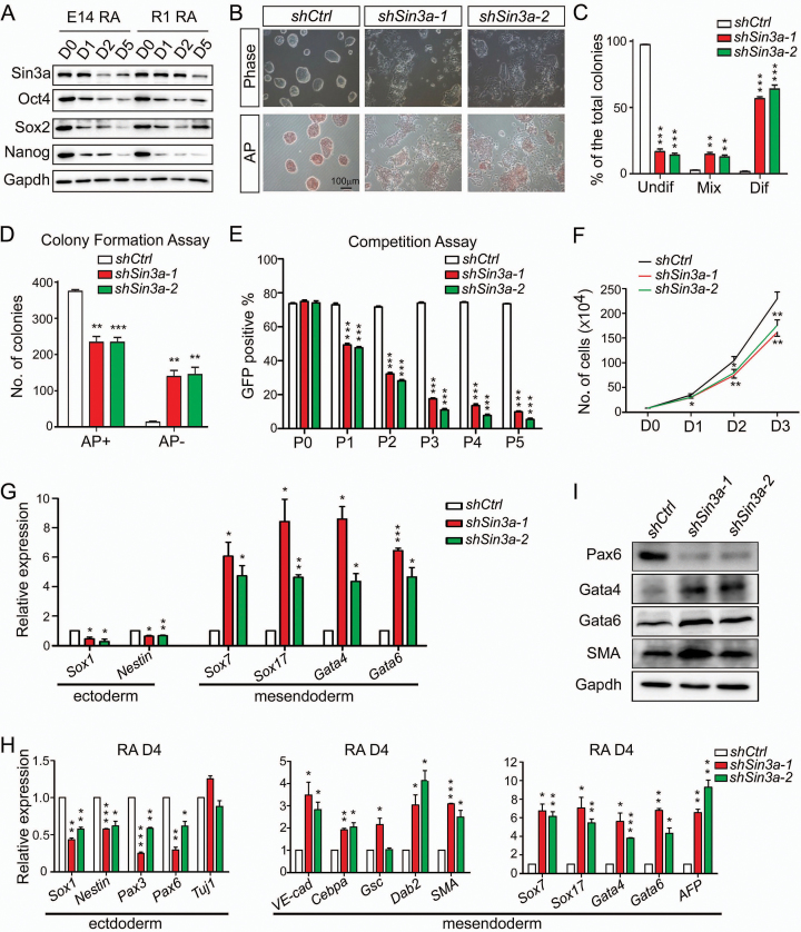
Knockdown of Sin3a impairs ESC self-renewal and skews differentiation toward mesendodermal fate. (A) Western blot analysis for Sin3a, Oct4, Sox2 and Nanog expression changes during 4 days of RA-induced ESC differentiation. (B) Cell morphology changes and AP staining after 4 days of Sin3a knockdown. (C) Statistical assay for the percentages of undifferentiated (Undif), mixed (Mix) and differentiated (Dif) ESCs described in (B). (D) Colony formation assay with AP staining following Sin3a knockdown. (E) Competition assay with a FACS plot showing the percentages of GFP positive (GFP+) ESCs that were respectively infected with lentivirus expressing shSin3a-1, shSin3a-2 or control shRNA. (F) Growth curve of ESCs following Sin3a knockdown. (G) RT-qPCR analysis of several early differentiation markers after 4 days of Sin3a knockdown under self-renewing conditions. (H, I) RT-qPCR (H) and western blot (I) analyses of various lineage markers at day 4 of RA-induced differentiation from Sin3a knockdown ESCs. Ctrl: control (and similarly hereafter). Data are representative of one experiment with at least three independent biological replicates. Data in (C–H) represent mean ± S.E.M. *P< 0.05, **P< 0.01, ***P< 0.001 versus the control, Student's t-test (n = 3).

