Figure 4.
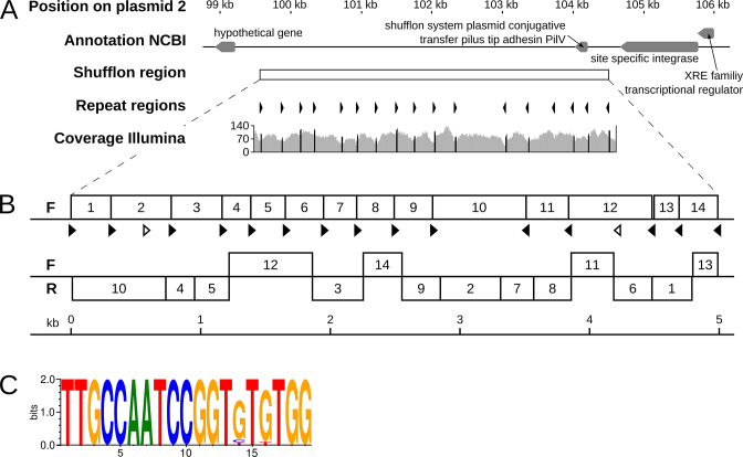
Overview of a shufflon region on plasmid 2. (A) The genomic region surrounding the shufflon region on plasmid 2 (top track) is shown along with gene annotations from NCBI (second track). The shufflon region is indicated as white box (third track), and the orientations of the inverted repeats are shown (as black arrows, track 4). The repeats are all pointing inwards to a position at around 102.5 kb. The coverage of mapped Illumina reads is shown (track 5). (B) Structure of the two major shufflon compositions identified. The upper version represents the sequence incorporated into the final assembly of P19E3. The lower variant exhibited extensive rearrangement of the shufflon locus (F: forward strand; R: reverse strand). (C) Consensus of inverted repeats of 19 bp length represented as a sequence logo (40).

