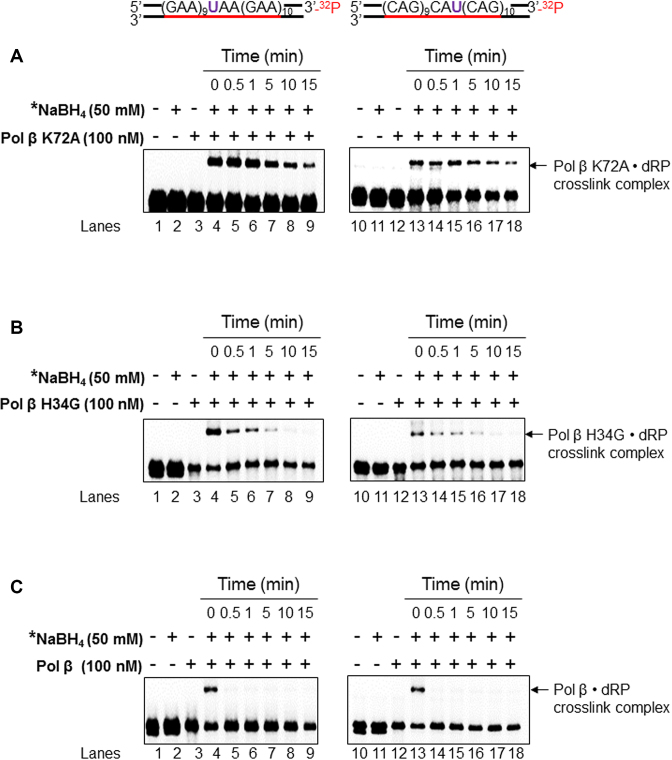
Figure 7.
The crosslink between the dRP lyase domain of wild-type and mutant pol β proteins and a dRP group. The formation of the pol β K72A•dRP crosslink complex (A), pol β H34G• dRP crosslink complex (B), and pol β• dRP crosslink complex (C) in TNRs, respectively was measured using the (GAA)20 (left panel) or (CAG)20 (right panel) substrates containing 5′- dRP as described in the Materials and Methods. Lane 1 indicates the uracil-containing substrate that was pre-incubated with UDG. Lane 2 indicates the pre-cut substrate in the presence of 50 mM NaBH4. Lanes 3 and 4 represent the reaction mixtures containing the pre-cut substrates and pol β K72A or H34G or pol β (wild-type) (100 nM) in the absence and presence of 50 mM NaBH4. Lanes 5 to 9 represent reaction mixtures that contained the substrates and pol β K72A or H34G or pol β (100 nM) in the presence of 50 mM NaBH4 with pre-incubation at the time points of 30 s, 1 min, 5 min, 10 min and 15 min. ‘*’ denotes 50 mM NaBH4 was added after pre-incubation with pol β K72A or H34G or pol β at various time intervals. Substrates were 32P-labeled at the 3′-end of the downstream damaged strand and are illustrated above each gel. Each experiment was repeated in triplicate. Only the representative gels are shown.

