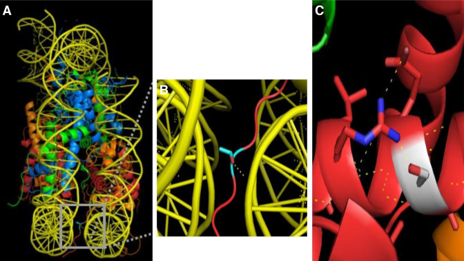
Figure 6.
Single amino acid substitutions in H2B affect nucleosome structure. (A) Threonine 33 of the H2B N-terminal tail (colored in cyan) is positioned between the DNA gyres of the nucleosome and hydrogen bonds to one strand of DNA. (B) Zoomed-in view of the region highlighted with a gray box in (A). Dashed yellow lines indicate hydrogen bonds identified by the PyMol polar contacts tool. (C) Serine 76 of H2B (shown in white) makes intra-chain interactions with the adjacent arginine. The hydrogen bond network thus formed between adjacent residues is shown with yellow dashed lines. Glycine 76 was replaced with serine using the PyMol mutagenesis tool. For all images, H3 is light blue, H4 is green, H2A is orange, and H2B is red. The crystal structure of the canonical nucleosome (PDB: 1AOI) was used for all images.

