
Figure 5.
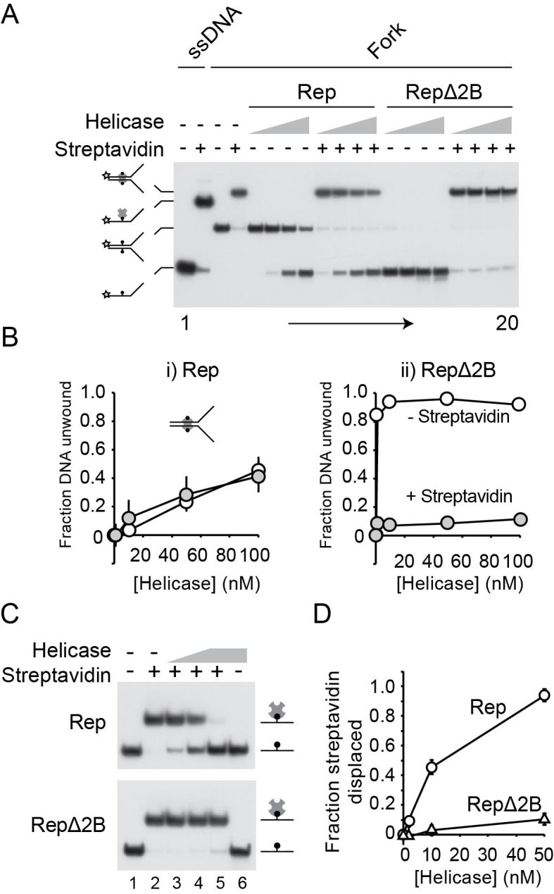
RepΔ2B is selectively inhibited by streptavidin-biotin complexes within the context of both ssDNA and dsDNA. (A) Native polyacrylamide gel of Rep- and RepΔ2B-catalysed unwinding of a forked DNA bearing biotin groups on both strands within the duplex portion of the fork (46). Lanes 1–4 contain markers indicating the positions of single-stranded and double-stranded DNA without and with bound streptavidin as indicated. Lanes 5–20 contain the products of unwinding of the forked DNA substrate in the absence and presence of streptavidin. The indicated helicase was present at 1, 10, 50 and 100 nM final concentration. (B) Quantification of unwinding of the biotinylated fork in the absence and presence of streptavidin. (C) Native polyacrylamide gel showing the impact of wild type Rep and RepΔ2B on a complex containing streptavidin bound to a biotin group within a single-stranded oligonucleotide. Lanes 1 and 2 do not contain helicase. Lanes 3 and 4 contain 2 and 10 nM whilst lanes 5 and 6 contain 50 nM helicase. (D) The degree of streptavidin displacement from the single-stranded oligonucleotide by wild type Rep and RepΔ2B.