Figure 5.
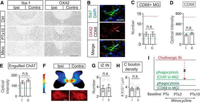
Systemic minocycline prevents microglial phagocytosis and rescues the downregulation of the cholinergic system after PTx. A, OX42, but not Iba-1, signal in microglia was substantially depressed in the IZ of the minocycline-treated spinal cord compared with that of untreated PTx10 spinal cord. B, Images of individual Iba-1+ microglial cells showed suppressed expression of OX42 and CD68 after minocycline treatment. C, Numbers of CD68+ phagocytic microglia in the medial IZ of minocycline-treated animals showing no increase. D, E, Optical intensity of CD68 and engulfed ChAT clusters in microglial cells in the medial IZ did not show the expected increase after injury. C–E, Mann–Whitney U test, C; p = 0.4676, n = 6 sections, D: p = 0.2443, E: p = 0.1029, n = 21–27 cells. The red dotted line indicates the mean value of PTx10 contralateral and the blue dotted line indicates the mean value of controls. F–H, Cholinergic IN distribution heat map and neuronal counting in the medial IZ showing a symmetric distribution pattern in the spinal cord of minocycline-treated animals, similar to that of control animals. The characteristic decrease in C bouton density after PTx also was blocked with minocycline treatment. G, H, Wilcoxon matched-pairs test, p = 0.5, n = 4. The color scale represents 0–4 cells/104 μm2. I, Schematic summary of changes in the cholinergic pathway and phagocytosis after minocycline treatment. With minocycline prevention of microglial phagocytosis (microglial ChAT/CD68), the cholinergic pathway (including INs and C boutons) was rescued from downregulation (broken red dotted arrow). Scale bars: A, 0.2 mm; B, F, 10 μm. See Table 1 for mean ± SEM values and Figure 5-1, for raw data. For all other statistical values, see Table 2.

