Figure 7.
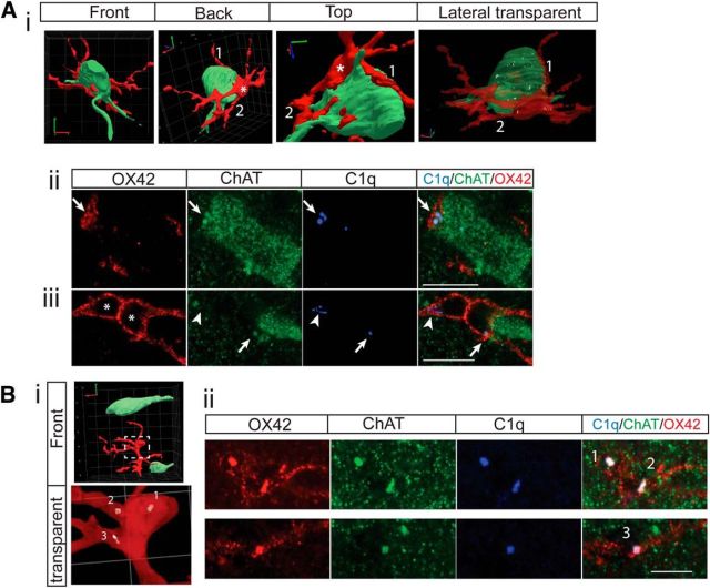
3D reconstruction of the interactions between microglial cells and cholinergic INs in the contralateral medial IZ. A, One ChAT+ IN and one microglial cell from a PTx3 animal. Ai, 3D reconstruction images show tight wrapping of the IN by a microglial cell from different angles. The semitransparent image shows numerous C1q clusters inside the cholinergic IN and microglia. Representative single optical images in Aii and Aiii show C1q+/ChAT+ clusters at the contact point of the neuron and microglial cell (arrows), indicating ongoing engulfment, endogenously expressed C1q in microglial cells (arrowhead), and the two chambers of the same microglial cell (*). B, Cholinergic INs and a microglial cell from a PTx10 animal. Bi, 3D reconstruction images showing little contact between the IN and the microglial cell. Transparent view shows dense C1q clusters inside the microglia, but none in neurons. Bii, Single optical images showing several C1q+/ChAT+ clusters inside microglia (1–3), indicating completed engulfment. Scale bar, 10 μm.

