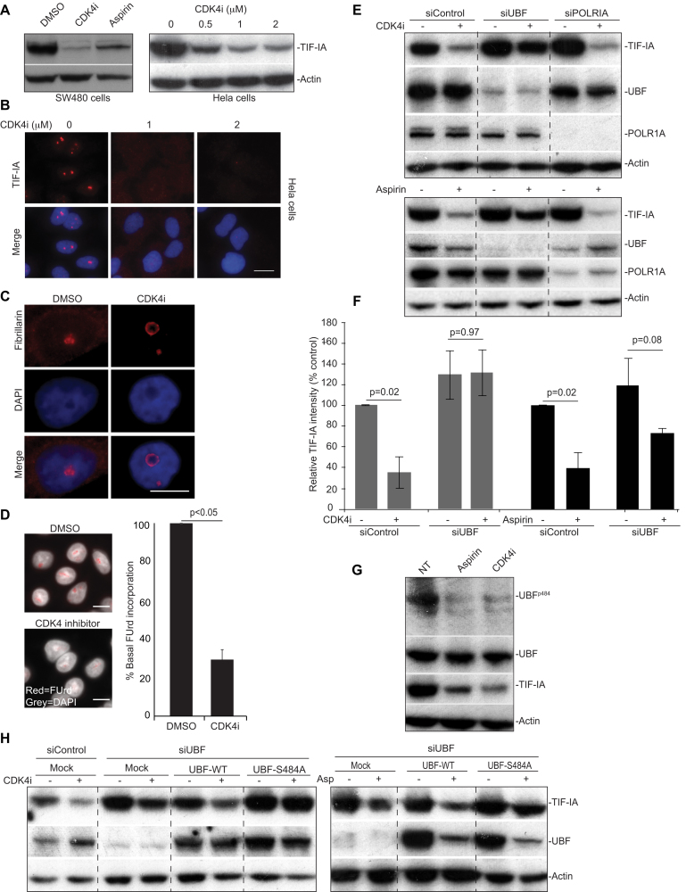
Figure 4.
A role for CDK4 and UBF S484 in TIF-IA degradation. (A–D) CDK4 inhibition induces degradation of TIF-IA and atypical changes to nucleolar structure. SW480 or Hela cells were treated with DMSO (carrier), aspirin (3 mM, 16 h) or the small molecule CDK4 inhibitor, 2-bromo-12,13-dihy-dro-indolo[2,3-a]pyrrolo[3,4-c] carbazole-5,7(6H)-dione (CDK4i, 2 uM or as indicated). (A) Anti-TIF-IA immunoblot performed on WCL. (B) Immunomicrographs (63×) demonstrating the levels and localisation of TIF-IA in Hela cells. (C) Immunomicrograph (63X) demonstrating re-localisation of fibrillarin in response to CDK4i in SW480 cells. (D) Left: Immunomicrographs (40×) depicting cells subjected to fluouridine (FUrd) run on assays. Right: Images were captured and analysed for FUrd incorporation using ScanR image analysis software. The results are presented as the percentage incorporation compared to control. The mean (±s.e.m.) of at least 1000 cells per experiment is shown. N=3 (E and F) SW480 cells were transfected with control, UBF or POLRIA siRNA. Forty-eight hours later cells were treated (+) with CDK4i (2 uM, 16 h), aspirin (3 mM 16 h) or the equivalent carriers (–). (E) Western blot analysis was performed with the indicated antibodies. (F) TIF-IA intensity (relative to actin) was determined for each condition using ImageJ analysis. Results are presented as the percentage relative TIF-IA compared to carrier treated, siControl. Mean (± s.e.m.) is shown for 2 (CDK4i) and 3 (aspirin) experiments. (G and H) Identification of a role for residue 484 of UBF. (G) SW480 cells were treated with aspirin and CDK4i as above. Western blot analysis was performed with antibodies to phosphorylated (UBF S484) and native UBF. (H) SW480 cells were transfected with control or UBF siRNA then either mock transfected or transfected with Flag-UBF-wild type (WT) or a phospho-mutant-flag-UBFS484A. Eight hours later, transfected cells were treated with CDK4i and aspirin (asp.) as above. Immunoblot was performed with the indicated antibodies. Scale bar = 10 μm. Actin acts as a loading control throughout. P values were derived using a two tailed Student's t test. See also Supporting Supplemental Figure S3.

