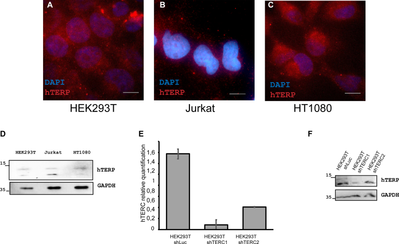
Figure 2.
hTERP is expressed in telomerase-positive cells. (A–C) Immunolocalization of hTERP in HEK293T (A), Jurkat (B) and HT1080 (C) cells; blue, DAPI-stained nuclei; red, hTERP; scale bars, 5 μm. (D) Western blot analysis of HEK293T, Jurkat and HT1080 cells for the presence of hTERP. GAPDH staining was used as a loading control. (E) Levels of hTERC in HEK293T shLuc (expressed shRNA targeted luciferase mRNA (negative control)), HEK293T shTERC1 and HEK293T shTERC2 (expressed shRNAs targeted hTERC) normalized to the hTERC level in HEK293T cells. Mean ± SD was calculated for three biological replicates. (F) Western blot analysis of HEK293T shLuc (expressed shRNA targeted luciferase mRNA (negative control)), HEK293T shTERC1 and HEK293T shTERC2 (expressed shRNAs targeted hTERC) for the presence of hTERP. GAPDH staining was used as a loading control.

