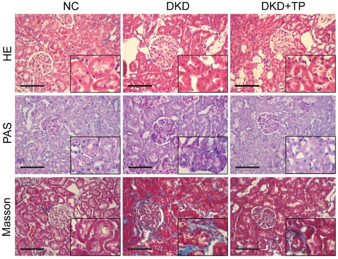
Figure 1.
Renal pathological changes in animal subjects. Representative images of hematoxylin and eosin (HE), periodic acid-Schiff (PAS) and Masson's trichrome (Masson) stained kidney sections (inset images indicate augmentative renal tubules). Original magnification is ×400. The scale bar represents 100 μm. NC: normal control; DKD: diabetic kidney disease; TP: triptolide.

