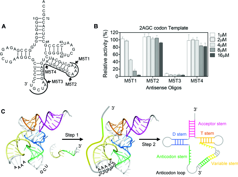
Figure 3.
Truncation analysis of M5-1 oligonucleotides. (A) Schematic representation of the M5-1 truncations. The black line shows the region of tRNASerGCU targeted by M5-1 oligonucleotide. M5T1 and M5T2 are truncated from 5′-end while M5T3 and M5T4 are truncated from 3′-end (indicated in the figure by the arrows). (B) Translational activity of 2AGC-codon eGFP-coding template in antisense oligo-treated E. coli lysate. The used concentrations of the oligonucleotides are coded by increasing shading of the graphs. The error bars represent standard deviations of at least two independent experiments. (C) Proposed mechanism of antisense oligonucleotide: tRNA interaction. The crystal structure of a tRNASec (PDB: 3W3S) was used to represent the E. coli tRNASerGCU. The loop regions are shown in grey while the stems are in colour. The nucleotides in the variable loop and the anticodon loop that are expected to be solution-exposed are marked. The antisense oligonucleotide is shown as an unstructured single-stranded sequence. The initial binding (Step 1) of the methylated antisense oligonucleotide to tRNASerGCU is proposed to initiate at the four consecutive adenosines of the variable loop facilitating duplex propagation (Step 2).

